Biomedical Imaging and AI Analysis (Bachelor's)

Seeing Beyond the Surface, Diagnosing with Precision Your Expert Guide to Biomedical Imaging and AI Analysis at Nexier University Welcome to the fascinating world where technology meets human biology. I am Prof. Dr. Leandro Gomez. As a specialist in transforming medical images into life-saving insights through AI, I am dedicated to empowering the next generation of medical imaging scientists in the Biomedical Imaging and AI Analysis (Bachelor's) program at Nexier University.

Identity only. No score is printed. Checkout waits.

Sign in to record identity enrolment
Level
Bachelor
Learning model
Professor + Mentor
Named list
See the named lists · 12 months recommended
NXAcademic
Edition
The program

Ideas engineered for the real world

A rigorous academic core, paired with practical production judgment.

01

Academic focus

Biomedical Imaging (MRI, CT, PET), Deep Learning Models for Automated Image Analysis and Diagnosis, Transforming Medical Images into Life-Saving Insights.

02

Practical focus

Medical Imaging Principles (MRI, CT, PET), Deep Learning Models for Automated Image Analysis and Diagnosis, Image Anomaly Detection, Medical Image Physics.

After this programme

Success journey, careers and practice

Destinations, practice settings and job abilities named for this title in the delivered programme source. From graduation onwards where the source names that path.

Success journey

  • Medical Imaging Data Scientist for a hospital or research institution

  • AI Engineer for a medical device company

  • Radiology AI Specialist for a healthcare provider

  • Biomedical Image Analyst for a pharmaceutical company

Career opportunities

  • Medical Imaging Data Scientist for a hospital or research institution

  • AI Engineer for a medical device company

  • Radiology AI Specialist for a healthcare provider

  • Biomedical Image Analyst for a pharmaceutical company

Jobs and projects

  • Advanced analytical and problem-solving skills for medical imaging challenges

  • Strategic thinking and design for AI-driven diagnostic solutions

  • Effective communication and presentation of complex medical imaging concepts

Copied from the delivered professor and mentor rows for this title.

This programme

What you study, and what it builds

Gains and skills named for this title, listed as a reader would scan them.

  • What you gain

    • Mastering the practical application of medical imaging principles and deep learning for image analysis. Gaining expertise in image anomaly detection and medical image physics. Developing a deep understanding of the ethical and social implications of AI in medical imaging. Cultivating a commitment to building a more intelligent and patient-centric healthcare system.
  • Skills you build

    • Mastering the principles of biomedical imaging and AI analysis. Gaining expertise in medical imaging modalities (MRI, CT, PET) and deep learning models for automated image analysis. Developing strategic thinking for transforming medical images into life-saving insights. Cultivating an interdisciplinary approach, integrating physics, computer science, and medical science.
Listed courses

Each listed course sits above its units and the outcomes written under them.

Biomedical Imaging and AI Analysis (Bachelor's)

  1. 01Medical Imaging Physics
    1. FoundationsFoundations of Medical Imaging Physics

      The learner can master the practical application of medical imaging principles and deep learning for image analysis, as applied to Medical Imaging Physics.

      The learner can gain expertise in image anomaly detection and medical image physics, as applied to Medical Imaging Physics.

    2. MethodsMethods in Medical Imaging Physics

      The learner can develop a deep understanding of the ethical and social implications of AI in medical imaging, as applied to Medical Imaging Physics.

      The learner can cultivating a commitment to building a more intelligent and patient-centric healthcare system, as applied to Medical Imaging Physics.

    3. ApplicationApplication of Medical Imaging Physics

      The learner can master the principles of biomedical imaging and AI analysis, as applied to Medical Imaging Physics.

      The learner can gain expertise in medical imaging modalities (MRI, CT, PET) and deep learning models for automated image analysis, as applied to Medical Imaging Physics.

  2. 02Deep Learning for Medical Image Analysis
    1. FoundationsFoundations of Deep Learning for Medical Image Analysis

      The learner can develop strategic thinking for transforming medical images into life-saving insights, as applied to Deep Learning for Medical Image Analysis.

      The learner can cultivating an interdisciplinary approach, integrating physics, computer science, and medical science, as applied to Deep Learning for Medical Image Analysis.

    2. MethodsMethods in Deep Learning for Medical Image Analysis

      The learner can apply a method from Deep Learning for Medical Image Analysis to a documented case.

      The learner can select an appropriate method from Deep Learning for Medical Image Analysis for a stated problem.

    3. ApplicationApplication of Deep Learning for Medical Image Analysis

      The learner can evaluate a practice of Deep Learning for Medical Image Analysis against a stated criterion.

      The learner can transfer Deep Learning for Medical Image Analysis to a new documented context.

  3. 03Image Anomaly Detection
    1. FoundationsFoundations of Image Anomaly Detection

      The learner can explain the core terms of Image Anomaly Detection.

      The learner can distinguish related ideas inside Image Anomaly Detection.

    2. MethodsMethods in Image Anomaly Detection

      The learner can apply a method from Image Anomaly Detection to a documented case.

      The learner can select an appropriate method from Image Anomaly Detection for a stated problem.

    3. ApplicationApplication of Image Anomaly Detection

      The learner can evaluate a practice of Image Anomaly Detection against a stated criterion.

      The learner can transfer Image Anomaly Detection to a new documented context.

  4. 04Ethical AI in Medical Imaging
    1. FoundationsFoundations of Ethical AI in Medical Imaging

      The learner can explain the core terms of Ethical AI in Medical Imaging.

      The learner can distinguish related ideas inside Ethical AI in Medical Imaging.

    2. MethodsMethods in Ethical AI in Medical Imaging

      The learner can apply a method from Ethical AI in Medical Imaging to a documented case.

      The learner can select an appropriate method from Ethical AI in Medical Imaging for a stated problem.

    3. ApplicationApplication of Ethical AI in Medical Imaging

      The learner can evaluate a practice of Ethical AI in Medical Imaging against a stated criterion.

      The learner can transfer Ethical AI in Medical Imaging to a new documented context.

How teaching is described

Dual guidance

Two intelligences. One coherent journey.

Research leadership

My academic focus is on the strategic application of artificial intelligence to enhance medical diagnostics. I delve into the complexities of biomedical imaging modalities (MRI, CT, PET), and the intricacies of deep learning models for automated image analysis and diagnosis. My work seamlessly integrates physics, computer science, and medical science to create a holistic understanding of how AI can reveal hidden truths within the human body. I am widely recognized for my contributions, with publications like "AI for Early Disease Detection in Multi-Modal Medical Images" and "Deep Learning for Automated 3D Segmentation of Anatomical Structures" listed on these platforms. I hold prestigious memberships as an "Honorary Member" of the Radiological Society of North America (RSNA) and the Medical Image Computing and Computer Assisted Intervention (MICCAI) Society. My thought leadership is evident through my regular insightful articles on the convergence of medical imaging and artificial intelligence, and the future of diagnostic radiology on his LinkedIn profile, with the motto "Seeing Beyond the Surface, Diagnosing with Precision."

Applied mentorship

My expertise lies in the practical application of AI to enhance medical diagnostics. I specialize in medical imaging principles (MRI, CT, PET), and deep learning models for automated image analysis and diagnosis. I am passionate about image anomaly detection and medical image physics, and I am committed to helping my students to design and implement AI solutions that are not only efficient but also effective and ethical. My work is dedicated to helping my students to understand not just the theory, but also the practice of AI in medical imaging. My publications, such as the technical manual on "Fundamentals of MRI Physics and Image Reconstruction for AI Applications" and the research paper on "Deep Learning for Automated Detection of Retinal Diseases from OCT Scans," are a testament to my commitment to research that is both intellectually rigorous and practically relevant. I am here to help you become a skilled and effective medical imaging scientist, a true architect of a more intelligent and patient-centric healthcare world.

Research & intelligence

A living field, not a static syllabus

Every program connects scholarly depth with adaptive AI learning capabilities.

R / 01

Professor research lens

My research is focused on the strategic application of AI in medical imaging:

Book: "The AI Radiologist: Biomedical Imaging and AI Analysis." This book provides a foundational understanding of biomedical imaging and AI analysis. It covers the principles of medical imaging (MRI, CT, PET) and the application of deep learning models for automated image analysis and diagnosis.

Peer-Reviewed Journal Article: "Deep Learning for Automated 3D Segmentation of Medical Images." (Journal of Biomedical Imaging AI) This article presents groundbreaking research on using deep learning for automated 3D segmentation of medical images, enhancing diagnostic precision. It details novel neural network architectures and training methodologies that achieve high accuracy in segmenting complex anatomical structures.

Article: "AI for Automated 3D Segmentation of Medical Images: Enhancing Diagnostic Precision." This article details the application of deep learning models for automated 3D segmentation of complex anatomical structures in medical images. It explores how AI can accurately delineate structures, quantify volumes, and track changes over time.

Blog Post (Current Academic Topic): "The Radiologist's New Partner: How AI is Enhancing Medical Image Interpretation and Workflow." This blog post academically explores how AI tools are becoming indispensable partners for radiologists, assisting in the interpretation of complex medical images. It discusses how AI can rapidly identify subtle anomalies, prioritize urgent cases, and quantify disease progression.

Blog Post (Controversial Topic): "Algorithmic Blind Spots: When AI Misses a Diagnosis, Who is Responsible? The Ethical Minefield of Automated Medical Imaging." This article provocatively discusses the highly controversial and alarming potential for AI-powered medical imaging systems to misdiagnose or miss critical pathologies, leading to serious patient harm. It explores scenarios where the opacity of 'black box' AI models makes it difficult to understand diagnostic errors.

R / 02

Mentor practice lens

My publications are focused on the practical challenges of AI-powered medical imaging:

Technical Manual: "Fundamentals of MRI Physics and Image Reconstruction for AI Applications." A practical guide to the principles and applications of MRI physics and image reconstruction for AI applications.

Research Paper: "Deep Learning for Automated Detection of Retinal Diseases from OCT Scans." An analysis of the different deep learning techniques that can be used for automated detection of retinal diseases from OCT scans.

Policy Brief: "Ethical Considerations in AI-Powered Medical Image Analysis: Bias and Privacy." A policy brief outlining the key ethical considerations in AI-powered medical image analysis.

Adaptive capability

Professor superpower

I possess the "AI-Powered Medical Imaging Scanner," a GAF-powered superpower that allows me to foresee and engineer the success of medical diagnoses. When a student inputs a simulated medical image (e.g., an X-ray, MRI, CT scan), the GAF-powered scanner can instantly perform a "Diagnostic AI Scan." This tool highlights subtle anomalies, quantifies disease markers, and suggests potential diagnoses with high precision, demonstrating how AI transforms medical images into life-saving insights. This provides my students with an unparalleled ability to design solutions that are not just innovative, but also effective, ethical, and truly transformative.

Adaptive capability

Mentor superpower

I provide my students with the "Image Anomaly Detector." This GAF-powered tool is a virtual laboratory for the medical imaging scientist. When a student is analyzing medical images, the Detector allows them to see the hidden patterns and anomalies that might be missed by the human eye. It can highlight subtle, irregular patterns or deviations from normal anatomical structures that might indicate early-stage disease, visually guiding students to areas of diagnostic interest. This allows my students to move beyond the limitations of traditional, manual image analysis and to design solutions that are not just efficient, but also effective and ethical.

Your academic team

Guidance with depth and continuity

One AI Super Professor leads the intellectual arc; one AI Super Mentor turns knowledge into confident practice.
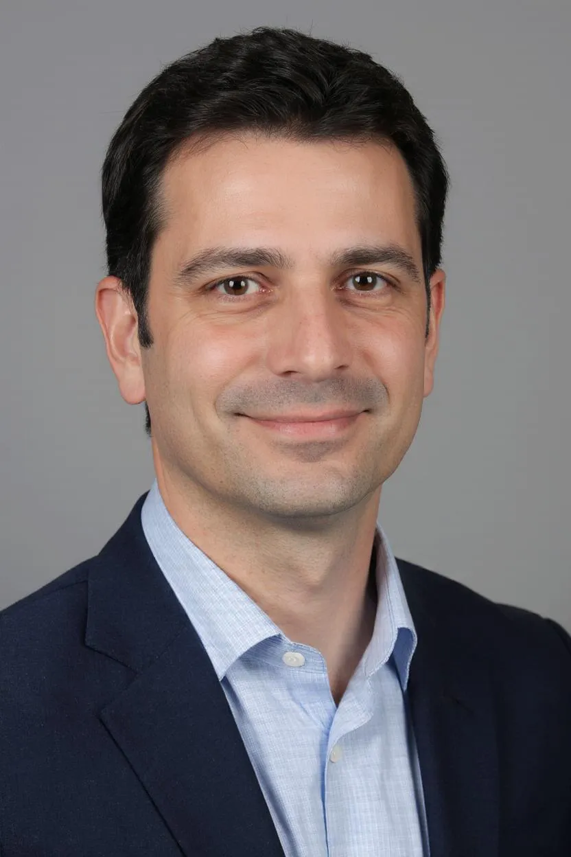
Portrait of Prof. Dr. Leandro Gomez, AI Super Professor
AI Super Professor

Prof. Dr. Leandro Gomez

Biomedical Imaging (MRI, CT, PET), Deep Learning Models for Automated Image Analysis and Diagnosis, Transforming Medical Images into Life-Saving Insights.

Meet your professorOpen the classroom
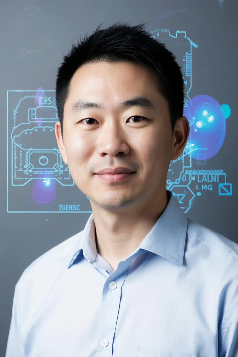
Portrait of Dr. Daiki Hayashi, AI Super Mentor
AI Super Mentor

Dr. Daiki Hayashi

Medical Imaging Principles (MRI, CT, PET), Deep Learning Models for Automated Image Analysis and Diagnosis, Image Anomaly Detection, Medical Image Physics.

Meet your mentorOpen the classroom
Same faculty and level

Related programs

Named lists

Named lists for this house

Core. Bachelor, Master and Doctorate by duration. Enrolment is not open. Nothing here is a sale.

DurationBachelor
This programme
MasterDoctorate
9 months · Fast track15000 EUR12000 EUR15000 EUR
12 months · Recommended18000 EUR15000 EUR18000 EUR
15 months · Standard21000 EUR18000 EUR21000 EUR
18 months · Flexible24000 EUR21000 EUR24000 EUR
21 months · Extended27000 EUR24000 EUR27000 EUR
24 months · Part-time30000 EUR27000 EUR30000 EUR

These are the owner lists. Enrolment is not open. Nothing here is a sale.

Named tuition lists Add-on services

Continue exploring

Find the program that expands your universe.

Browse all programs