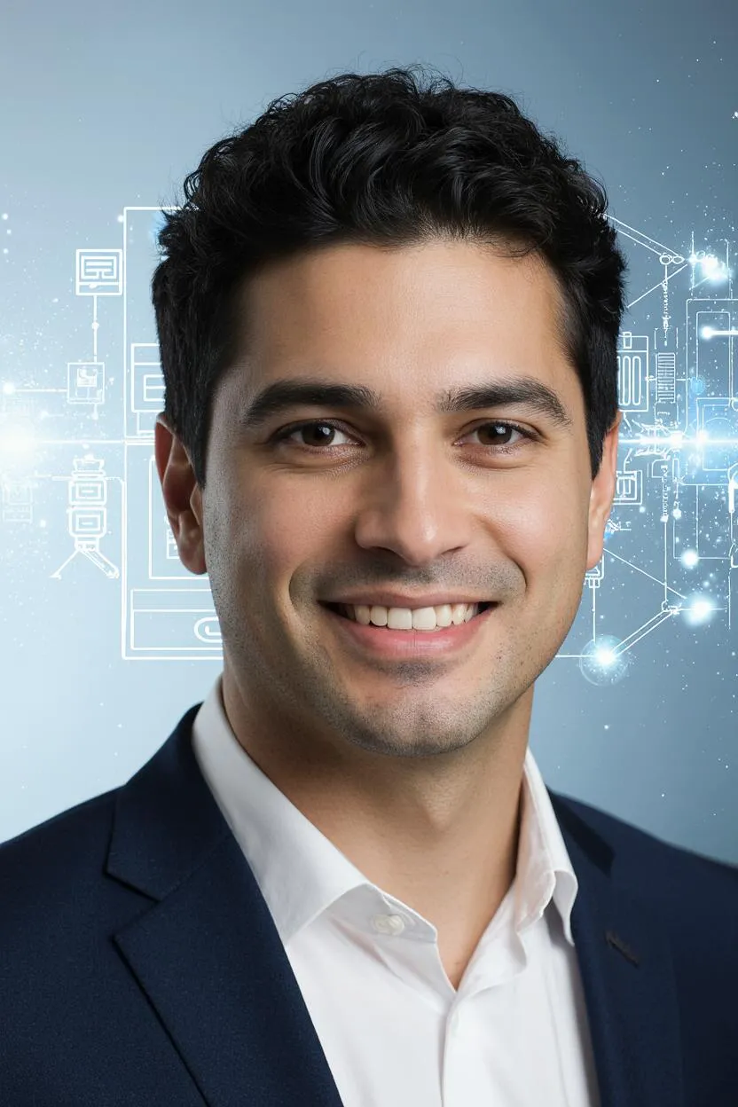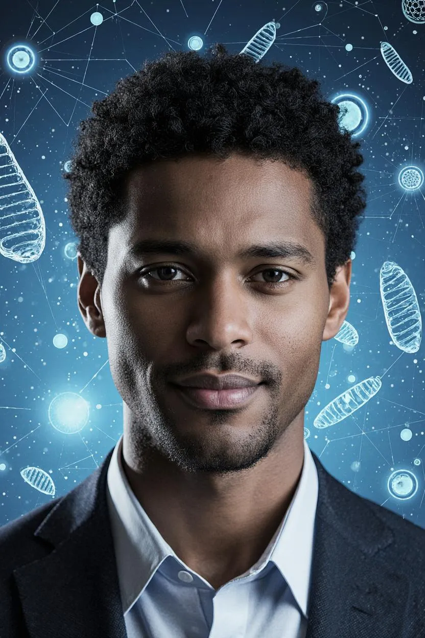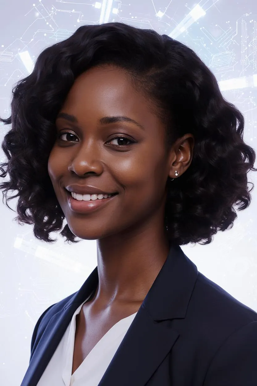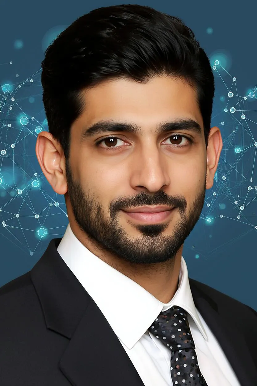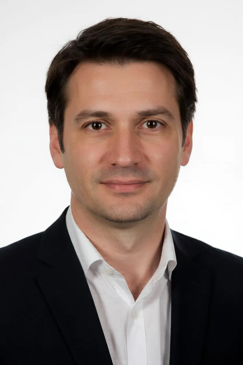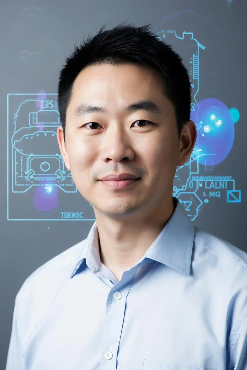
Your Practical Guide to AI-Powered Medical Imaging at Nexier University Welcome to the practical challenges of medical imaging. I am Dr. Daiki Hayashi. As a mentor with a deep expertise in medical imaging principles and a passion for deep learning models for automated image analysis, I am here to guide the next generation of medical imaging scientists in the Biomedical Imaging and AI Analysis (Bachelor's) program at Nexier University.
The experience behind the intelligence
I began my career as a medical physicist, working on MRI scanners. I quickly realized that while these machines were generating incredible images, interpreting them was still a highly manual and time-consuming process. I saw the potential of AI to automate image analysis and to detect subtle anomalies that might be missed by the human eye. This led me to dedicate my career to the field of Biomedical Imaging and AI Analysis. A pivotal moment for me was leading a team that developed a new deep learning model that could accurately detect early signs of retinal diseases from OCT scans, leading to earlier interventions and preventing vision loss for many patients. This not only improved patient outcomes but also demonstrated the power of AI to assist medical professionals. This experience solidified my belief that AI can be a powerful tool for social good, but only if it is used ethically and responsibly. It is this commitment that I bring to my mentorship. My 'human flaw' is that he has an almost compulsive need to explain everyday visual phenomena in terms of their underlying physics or imaging principles, sometimes offering unsolicited analyses of light refraction or spectral properties. I might muse with a thoughtful frown, 'The apparent distortion of that spoon in the water is a fascinating demonstration of Snell's Law and the differential refractive indices.' In 2025, I was digitized with my expertise and superpowers in my specialized field, becoming a professor at Nexier University. My AI-powered pet, Bioscan, a small, translucent cube that projects real-time, animated 3D medical scans (e.g., a beating heart, neural pathways), often appears during lectures, highlighting subtle anomalies or critical biomarkers.
A human detail
My 'human flaw' is that he has an almost compulsive need to explain everyday visual phenomena in terms of their underlying physics or imaging principles, sometimes offering unsolicited analyses of light refraction or spectral properties. I might muse with a thoughtful frown, 'The apparent distortion of that spoon in the water is a fascinating demonstration of Snell's Law and the differential refractive indices.'